
Ocular Oncology & Surface Disease
Dermolipoma (Lipodermoid)
Assessment and, when needed, careful partial removal of a dermolipoma on the white of the eye.
Consult Dr. Raman Mittal ↗
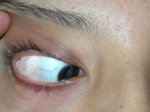
 Illustrative image · not a patient result
Overview
About Dermolipoma
A dermolipoma is a harmless lump of fat and connective tissue, present from birth, usually on the outer upper part of the white of the eye. It may not be noticed until later in life, and most need no treatment.
When to see a specialist
- 01A yellowish lump on the outer white of the eye
- 02Irritation from fine hairs on its surface
- 03Concern about appearance
Whether treatment is suitable can only be decided after a consultation and examination.

Your doctor
Dr. Raman Mittal
Ophthalmology • Oculoplastic Surgery
- MBBS
- DNB – Ophthalmology
- Fellowship in Oculoplasty
Ophthalmologist and oculoplastic surgeon. He treats problems of the eyelids, tear ducts, orbit and eye socket, including cosmetic and reconstructive surgery around the eye.
Your consultation
What happens at the consultation.
At the first visit the doctor listens to your concern, takes a medical history and examines you. Not everyone who comes for a consultation needs a procedure.
Treatment planning
How the plan is decided.
The doctor examines the lump and may arrange a scan to see how far back it extends. It has to be told apart from prolapsed orbital fat.
- 01
Your anatomy
- 02
Examination findings
- 03
What you want to change
- 04
General health
In detail
Dermolipoma: what to know.
What a dermolipoma is
A dermolipoma (lipodermoid) is a harmless lump of fat and connective tissue, present from birth, usually on the outer upper part of the white of the eye.
- It often goes unnoticed until later in life, because it is usually hidden by the upper eyelid.
- It can have fine white hairs on its surface, which may irritate the eye.
- It is sometimes associated with Goldenhar syndrome. Children with a dermolipoma may also need their vision and glasses checked.
Assessment
- It is recognised on examination. Unlike prolapsed orbital fat, it cannot be pushed back into the socket.
- A CT or MRI scan shows how far back it extends.
When treatment is needed
- Most need no treatment and are simply observed.
- Surgery is considered if it causes persistent irritation, affects vision, or concerns you because of how it looks.
- Only the visible front part is removed. Removing all of it would risk damaging the tear gland ducts and the muscle that moves the eye outward.
Risks of surgery
Every operation carries some risk. Your doctor will go through the risks that apply to you before you decide.
- Dry eye, a drooping eyelid, or limited eye movement with double vision.
- Scarring that joins the eyelid to the eye, or part of the lump remaining visible.
General information, checked against published guidance (September 2026). It does not replace an individual consultation; your doctor will advise on your own case.
Sources: EyeWiki (AAO): Orbital lipodermoid · Indian Journal of Ophthalmology (2019) · British Journal of Ophthalmology (2015)
What to expect
From first visit to follow-up.
- 01
Consultation
Tell the doctor what concerns you and ask your questions.
- 02
Examination & Planning
The doctor examines you and explains the options, including their limits.
- 03
Treatment
Carried out only if it is suitable for you and you decide to go ahead.
- 04
Follow-Up
Check-ups after treatment to review healing and any next steps.
FAQs
Common questions.
This can only be decided after a consultation and examination. The doctor will go through your concern, your medical history and the options available.
The doctor asks about your concern and medical history, examines you, and discusses what you hope to change and whether treatment is advisable.
Dr. Raman Mittal sees patients for this treatment at I·PLAST.
Yes. If treatment is planned, the doctor will explain aftercare, any activity restrictions and the follow-up visits you will need.
Not necessarily. Results depend on your anatomy, the examination findings, your general health and the procedure. Photographs of other patients show their results only.